Temporomandibular joint (TMJ) Disorders.
The i-CAT™ provides multi-planar or three-dimensional images of the condyle and surrounding structures to facilitate analysis and diagnosis of bone morphology, joint space and dynamic function, critical keys to providing appropriate treatment outcomes in patients with TMJ signs and symptoms.
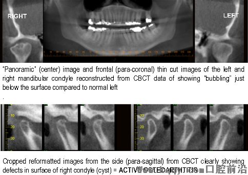
Degenerative Joint Disease (DJD or OA[osteoarthritis])
The following patient presented with limited opening of the jaw, pain in the right side and a swing (deviation) to the right with opening.
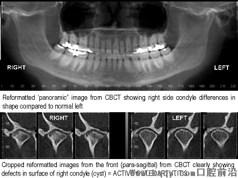
Another patient (below) presented with slow progressive changes in the bite of the teeth and was assessed prior to jaw (orthognathic) surgery. The changes in the TMJ are suggestive of active degenerative joint disease – an important consideration before such surgery.
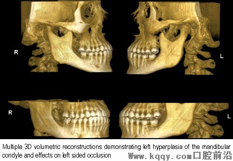
Developmental Anomalies of the Condyle
This patient presented with a changing bite located on the right after orthodontic treatment. The cause for this is easily determined by 3D reconstructed volumetric renderings showing substantial left condylar hyperplasia.
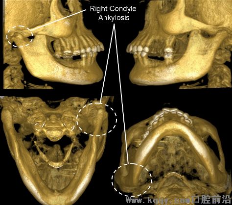
Ankylosis of the Condyle
This patient presented with progressing limited opening of the jaw. CBCT 3D volumetric reconstructions demonstrate a deformity on the right consistent with fusion (ankylosis) of the condyle to the skull.
Dental implants.
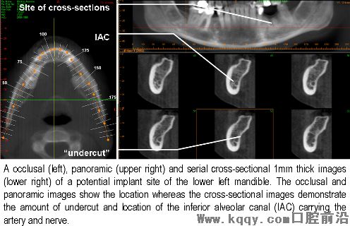
Cross-sectional imaging is crucial to the assessment of potential implant sites by providing information on the available bone height and width as well as assisting in implant angulation. The i-CAT™ provides cross-sectional images of the alveolar bone and accurately depicts vital structures such as the inferior alveolar dental nerve canal (IAC) in the mandible or maxillary sinus in the maxilla contributing to more accurate assessment and correct placement.
Examples of i-CAT™ imaging for implant assessment
One component of the successful placement of dental implants depends on the adequate assessment of the available height and width of alveolar bone at proposed implant sites. CBCT provides images at life-size which are familiar to surgeons (e.g. “panoramic”) and thin cuts at appropriate locations.
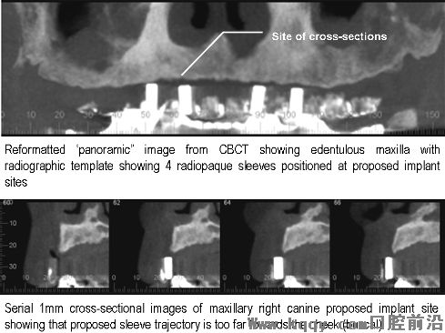
Another example of the assessment of a potential implant site in the lower left jaw (mandible) of a patient with a recent extraction.
Localization of teeth
The inherent high resolution of the i-CAT™ ensures anatomic accuracy in the localization of impacted teeth as well as the assessment of root resorption or fusion (ankylosis).
Location of impacted canines
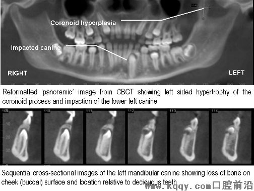
This young patient was referred from an orthodontist because the eruption of the left mandibular (lower jaw) canine was delayed as compared with the other side. The patient also had difficulty in opening. The location of the canine was easily determined however it was also shown that the left coronoid process was markedly elongated and was the most probable cause for the limited opening.
Location of Supernumerary teeth
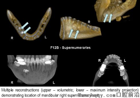
This patient presented with a request for imaging to provide accurate localization of supernumerary teeth in the lower right jaw prior to surgery. Cross-sectional images (not shown) were also used to determine if the unerupted teeth were stuck (ankylosed) in bone or if they had caused resorption of the roots of the erupted teeth.
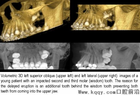
Delayed Eruption of teeth
This patient was referred for CBCT assessment because the maxillary left posterior molar teeth had failed to erupt. While conventional radiography showed multiple unerupted teeth, CBCT provided the accurate location of all teeth prior to surgical removal.
Assessment of pathology.
The inherent high resolution of the i-CAT™ ensures anatomic accuracy and assessment of all bony pathology in the maxillofacial region.
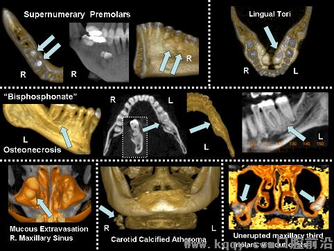
Evaluation of various anomalies
The montage below shows how various CBCT reconstructions can be used to visualize conditions of the jaws including; additional teeth (supernumeraries) (upper left); bony growths on the tongue (lingual) side of the mandible (upper right); thickening (osteosclerosis) and alteration of the mandibular bone in a patient on bisphosphonate therapy (center); soft tissue conditions (mucous extravasation cyst) of the right maxillary sinus (lower left); carotid artery calcifications (lower center) and; unerupted maxillary third molars and their relationship to the maxillary sinus (lower right).
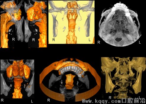
Location of calcifications in the neck.
Numerous calcifications can occur in the head and neck region. It is important to identify the location of these calcifications as this can aid in distinguishing benign (e.g. tonsiloliths, lymph nodes, salivary gland stones) from potentially significant calcifications of the arteries (as carotid artery calcifications) or veins (e.g phleboliths) This patient presented with bilateral calcifications adjacent to the lower jaw. The images shown her clearly demonstrate the calcifications as a series of small concresences immediately adjacent the oro-pharyngeal airway – this appearance is consistent with bilateral tonsiloliths.
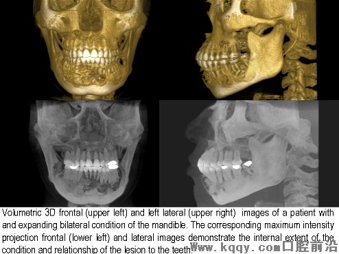
Benign Expansile Conditions
CBCT is capable of demonstrating the location, size, shape extent and full involvement of pathology of the upper or lower jaws.
3D imaging / stereolithographic modeling.
Data from the i-CAT™ can be reformatted to produce 3D images for the surgeon. This is important for patients requiring major reconstructive surgery. These images can be converted into models which assist the surgeon prior to the operation, reducing anesthetic cost and operating time.
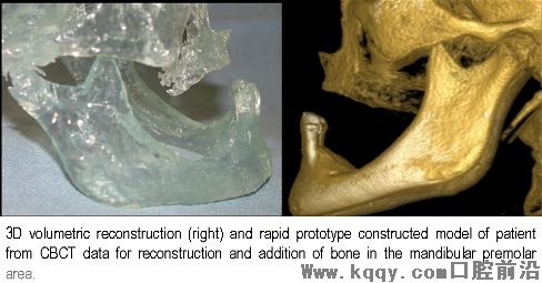
Biomodel Fabrication for Implant Treatment Planning
This patient had severe atrophy of the mandible bilaterally in the premolar area. Accurate plastic models (left) were fabricated from 3D models (right) to assist planning for block bone supplementation (augmentation) of the available ridge prior to implant placement.
Pre-Operative Evaluation
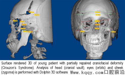
Craniofacial anomalies
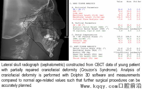
Full height scans of patients with craniofacial anomalies can be analyzed using additional software such that precise measurements of the skull and facial bones can be performed. These measurements compared to normal age-related values such that further surgical procedures can be accurately planned. Analysis can be performed both on 3D reconstructions (upper figure) and on conventional images (lower figure) – both generated from CBCT data.
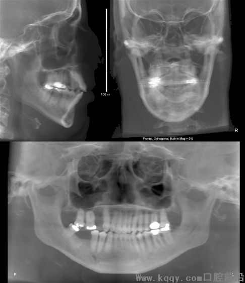
Dento-Maxillofacial Deformities / Asymmetry
This patient presented with an asymmetry of one side of the face. CBCT data can be reformatted to provide multiple conventional “standardized” images such as the lateral cephalometric (upper left), frontal cephalometric or posterior anterior (PA) (upper right) and panoramic (lower) projections.
Post operative evaluation
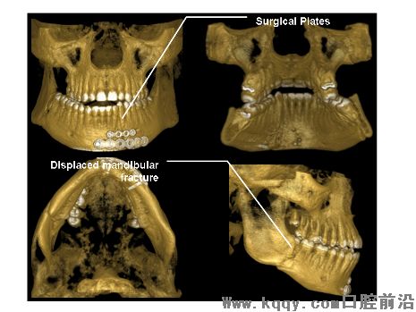
Trauma
This patient was referred for evaluation of the success of surgical plating of an anterior mandibular fracture. It was also discovered that the patient had a displaced fracture of the right mandibular body.
Orthognathic Surgery
This patient presented with some degree of pain after 12 weeks of orthognathic surgery to the upper and lower jaw to correct a severe facial deformity. Post-operative assessment of the patient was warranted to make sure that there was adequate healing and no movement or infection of the surgical plates.
相关阅读:
 发表评论用户评论